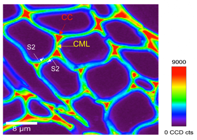
Figure 1. Chemical imaging of lignin in stem secondary xylem cells of P. trichocarpa by confocal Raman microscopy (Schmidt et al. 2009. Planta, 230:589-597). Lignin signal intensity is the strongest in CC, followed by compound middle lamella (CML). Lignin is also distributed in the S2 layer of the cell wall, with a higher concentration in the outer wall (towards CML).


